Fig. 3
OPTICS: MAGNIFICATION: DATE OF IMAGE: SUBMITTER COMMENTS:
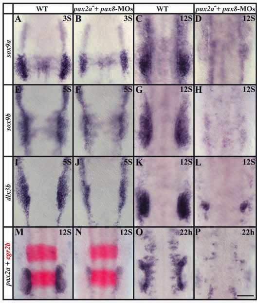
Fig. 3 Pax2a and Pax8 are required for maintenance of otic cell fates. Pax2a and Pax8 are required together for preotic expression of sox9a (A-D), sox9b (E-H) and pax2a (M-P) but not of dlx3b (I-L). Cells of the presumptive otic placode express sox9a in wild-type embryos at the three-somite stage (A) at higher levels than in pax2a– mutants after pax8-MOs injection (B). At the 12-somite stage, sox9a is expressed throughout the otic placode in wild-type embryos (C) but no otic sox9a expression can be detected in pax2a– mutants depleted of Pax8 (D). (E-H) The sox9a duplicate, sox9b, shows similar behavior. In wild type at the five-somite stage, sox9b is expressed in the preotic region and neural crest (E). The neural crest expression is unaffected in pax2a– mutants injected with pax8-MOs, but expression in the preotic domain is reduced (F). At the 12-somite stage, sox9b is expressed strongly in the otic placode in wild-type embryos (G) but is absent in pax2a– mutants injected with pax8-MOs (H). dlx3b expression is strong in cells of the future otic placode in wild-type embryos (I) at the five-somite stage and this domain is smaller but still recognizable in pax2a– mutants after pax8-MOs injection (J). At the 12-somite stage, dlx3b is expressed throughout the otic placode in wild-type embryos (K) but in pax2a– mutants with a knockdown of Pax8, only a few residual cells express dlx3b (L). pax2a expression is strong in the otic placode of wild-type embryos at the 12-somite stage (M) but expression is severely reduced in pax2a– mutants after pax8-MOs injection (N). At 22 h, when the otic vesicle has formed in wild-type embryos, pax2a expression is restricted to the ventromedial region (O) but is completely absent in pax2a– mutants depleted of Pax8 (P). Expression of egr2b (red) in rhombomeres 3 and 5 is unchanged in pax2a– mutants after pax8- MOs injection (N) in comparison with uninjected wild-type embryos (M). Dorsal views, anterior towards the top. Scale bar: 120 μm.
| Preparation | Image Form | View | Direction |
| not specified | still | not specified | not specified |

