
Fig. 3
- ID
- ZDB-FIG-240717-24
- Publication
- Chen et al., 2024 - Epidermal growth factor-like domain 7 drives brain lymphatic endothelial cell development through integrin αvβ3
- Other Figures
- All Figure Page
- Back to All Figure Page
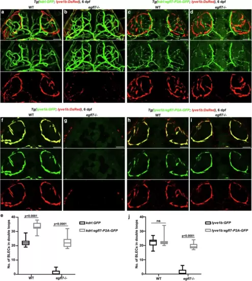
egfl7 regulates brain lymphatics development either autonomously or non-autonomously.a?d In the stable transgenic line Tg(kdrl:egfl7-p2A-GFP), the replenishment of Egfl7 in BVs can rescue the absence of BLECs in the egfl7 mutant (d) in contrast to the mutant under the Tg(kdrl: GFP) transgenic line (b). For WT, overexpression of Egfl7 in the BVs results in increased BLECs emerge in the bilateral loop over the brain (c) compared to the WT under the Tg(kdrl:GFP) (a). e The statistics show the number of BLECs per double loops in WT and egfl7 mutant under Tg(kdrl: GFP) and Tg(kdrl:egfl7-p2A-GFP) transgenic lines (n = 24 embryos, 2-way ANOVA. Box plots show the five-number summary of a set of data: including the minimum score (shown at the end of the lower whisker), first (lower) quartile, median, third (upper) quartile, and maximum score (shown at the end of the upper whisker)). f?i In contrast to the mutant under Tg(lyve1b: GFP) transgenic line (g), the replenishment of Egfl7 in LECs under Tg(lyve1b:egfl7-p2A-GFP) is able to re-form the BLECs bilateral loop in the egfl7 mutant (i). And overexpression of Egfl7 in WT lymphatics shows the BLECs are unaltered (f, h). j The statistics show the number of BLECs per double loops in WT and egfl7 mutant under Tg(lyve1b: GFP) and Tg(lyve1b:egfl7-p2A-GFP) transgenic lines (n = 24 embryos, 2-way ANOVA. Box plots show the five-number summary of a set of data: including the minimum score (shown at the end of the lower whisker), first (lower) quartile, median, third (upper) quartile, and maximum score (shown at the end of the upper whisker)). Scale bar, 50 ?m. |