
Fig. 8
- ID
- ZDB-FIG-240701-32
- Publication
- Pollitt et al., 2024 - Llgl1 mediates timely epicardial emergence and establishment of an apical laminin sheath around the trabeculating cardiac ventricle
- Other Figures
- All Figure Page
- Back to All Figure Page
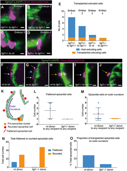
Cell-autonomous analysis of Llgl1 requirements in epicardial colonisation and cardiomyocyte extrusion. (A-D) Live light-sheet z-slices through the ventricle of Tg(myl7:LifeAct-GFP) transgenic embryos with rhodamine-dextran-labelled donor myocardial cells (magenta) at 76 hpf. Scale bars: 15 μm. (E) Quantification of host extruding cells and transplanted extruding cells in the four embryos with extruding transplanted CMs. (F-J) Live light-sheet z-slices through the ventricle of Tg(myl7:LifeAct-GFP) transgenic embryos with rhodamine-dextran-labelled donor epicardial cells (magenta) at 76 hpf. Scale bars: 15 μm. Transplanted epicardial cells can be flattened (white arrow, F,G,I) or rounded (yellow arrow, H,J). Epicardial cells can be seen surrounding an extruding CM (blue arrowhead, G). (K) Schematic depicting classification of epicardial cells and outer curvature. (L) Quantification of transplanted flattened mature epicardial cell number. Each dot represents a single embryo. (M) Quantification of number of epicardial cells on outer curvature. Each dot represents a single embryo. Data in L and M are mean±s.d. (N) Categorisation of total epicardial cells as flattened (mature) or rounded (immature). Flattened mature epicardial cells are significantly associated with wild-type donors (Fisher's exact test: P=0.0002). (O) Proportion of epicardial cells associated with the outer curvature. Wild-type transplanted epicardial cells are significantly more likely to be positioned on the outer curvature (Fisher's exact test: P=0.0252). |