|
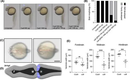
CRISPR/Cas9 dre-ca8 F0 knockout impairs early brain development. (A) Lateral view of 24 hpf zebrafish embryos (midbrain-hindbrain boundary indicated by purple arrowheads) and (B) quantification demonstrating altered development of the forebrain, midbrain, and hindbrain in ca8 crispant embryos. (C) ca8-crispants also present marked abnormal brain ventricle formation at 24 hpf. (D) Schematic illustration of the normal morphology of zebrafish brain ventricles at 24 hpf. (E) Quantification of abnormal brain ventricle formation observed at 24 hpf in dre-ca8 crispant embryos. Significance determined by Mann-Whitney tests. Scale bar, 250 ?m. **P < 0.01, ***P < 0.001, ***P < 0.0001. F, forebrain; M, midbrain; H, hindbrain; cb, cerebellum, see Pose-Méndez et al;16 dpf, days post fertilization; hpf, hours post fertilization [Color figure can be viewed at wileyonlinelibrary.com]
|

