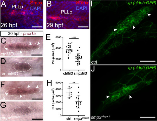
Smpx-deficiency causes the decrease in the PLLp size. (A,B) Immunofluorescence with the anti-Smpx antibody unveiling the localization of the protein in the PLLp at 26 and 29 hpf. (C,D) In-situ hybridization with a prox1a probe to compare the PLLp size (white-dotted encircled areas) in 30 hpf ctrlMO and smpxMO; the PLLp of Smpx-deficient embryos appears smaller in size; the typical rosette-like structures present in the ctrlMO sample (white arrowheads in C) are not clearly distinguishable in the smpxMO sample. (E) Size quantification in ctrlMO and smpxMO showing a drastic decrease in the PLLp area following the downregulation of smpx. (F,G) In-situ hybridization with a prox1a probe to compare the PLLp size (white-dotted encircled areas) in 30 hpf ctrl and smpxcrispant embryos; the PLLp of the smpxcrispant embryos appears smaller in size and the typical rosette-like structures present in the ctrl sample (white arrowheads in F) are not clearly distinguishable in the smpxcrispant preparation. (H) Size quantification in ctrlMO and smpxcrispant showing a drastic decrease in the PLLp area following the ablation of smpx. (I,J) Z-stack confocal images of the PPLp at 30 hpf in tg (cldnb:GFP) embryos; the shape of the epithelial rosettes in control embryos are lost in smpxcrispants. (A,B) Confocal Z-stacks taken from whole mount embryos; lateral views, anterior to the left. Embryos were counterstained with DAPI to visualize cell nuclei. (E,H) Unpaired t-test was performed to assess statistical significance by setting p ≤ 0.05 (*), p ≤ 0.01 (**), p ≤ 0.001 (***) and p ≤ 0.0001 (****) as significant. (E) ctrlMO n = 22, smpxMO n = 18. (H) ctrl n = 10, smpxcrispant n = 13. Scale bars are 50 µm.
|
