
Fig. 1
- ID
- ZDB-FIG-240314-8
- Publication
- Chiang et al., 2023 - The Role of MAPRE2 and Microtubules in Maintaining Normal Ventricular Conduction
- Other Figures
- All Figure Page
- Back to All Figure Page
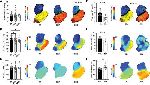
mapre2 loss of function leads to decreased ventricular conduction and voltage-gated sodium channel (NaV) function. A through C, Voltage mapping of hearts isolated from wild-type (WT) vs heterozygous (HET) and homozygous (HOMO) mapre2 knockout (KO) zebrafish. In HOMO hearts, there was a nonsignificant decrease in ventricular conduction velocity (CV; A), a significant decrease in ventricular maximum upstroke velocity of the action potential (Vmax; dV/dt; P=0.0121 vs WT; B), and a nonsignificant increase in ventricular action potential duration (APD; C). There was also a significant decrease in ventricular Vmax in HET hearts compared with WT (P=0.0363). Multiple comparisons were done using the Dunnett test if 1-way ANOVA was significant (P=0.0147 for Vmax in B). D through F, Voltage mapping of hearts isolated from control (CTL) vs mapre2 morpholino (MO)-injected larvae. In MO hearts, there was a significant decrease in ventricular CV (unpaired t test, P<0.0001; D) and Vmax (unpaired t test, P<0.0001; E) and a significant increase in ventricular APD (Mann-Whitney U test, P=0.0021; F). All hearts (represented by dots) were isolated from 5-dpf zebrafish larvae. The dotted squares in A and D reflect the main ventricular area in the hearts from which the parameters were measured. APD was measured at 80% repolarization while the hearts were paced at 100 bpm. |