|
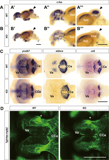
Defects in cerebellar structure and function in znf536 KO zebrafish. A, B”’ Reduced neuronal activity in KO zebrafish monitored by c-fos expression after novel tank assay, especially in the cerebellum (arrowheads). n = 3 for WT and n = 3 for KO. C Expression of Purkinje cell markers (aldoca, pvalb7, ca8) were specifically affected in the Va region. n = 3 for WT and n = 3 for KO for each marker. D Confocal images of myelinated bundles in Tg[mbp:mgfp]::znf536 KO brain, focused in the Va region. A unique circular structure (asterisk) was identified within the Va. n = 2 for WT and n = 2 for KO. CCe corpus cerebelli, Dm medial zone of the dorsal telencephalic area, TeO optic tectum, Va valvular cerebelli. Scale bars: 500 µm.
|