FIGURE

Figure 2

ID
ZDB-FIG-240109-65
Publication
Derrick et al., 2023 - Functional analysis of germline VANGL2 variants using rescue assays of vangl2 knockout zebrafish
Other Figures
All Figure Page
Back to All Figure Page
Figure 2

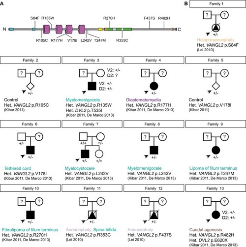
Collation of clinical data relating to historical VANGL2 variants. (A) Primary structure of human VANGL2 (UniProt: Q9ULK5) [116, 117] showing known functional domains and location of VUS. Two N-terminal serine/threonine motifs (cyan [78]), 4 transmembrane domains (magenta, [98]), TGN (trans-Golgi network) sorting motif (yellow [118]), the Dishevelled and Prickle binding region (green [119–121], which overlaps with two ubiquitinoylation sites, (orange [122], a VCP interacting motif (mauve, [122])) and a nuclear localization signal (white [123]). Two further ubiquitinoylation sites (orange) and the Type I PDZ domain (blue, [124]) are present at the C-terminal end. (B) Clinical pedigrees, major developmental defects and original references relating to previously reported missense mutations in coding sequence of human VANGL2. Squares, circles, diamonds, triangles represent male, females, unknown gender, and foetuses, respectively. Arrowheads denote proband. Diagonal line denotes deceased. Open symbols: healthy, black symbols: affected. V2: VANGL2 genotype, D2: DVL2 genotype. Question mark denotes DNA unavailable. Het.: heterozygous.

Expression Data

Expression Detail
Antibody Labeling
Phenotype Data

Phenotype Detail
Acknowledgments
This image is the copyrighted work of the attributed author or publisher, and ZFIN has permission only to display this image to its users. Additional permissions should be obtained from the applicable author or publisher of the image. Full text @ Hum. Mol. Genet.