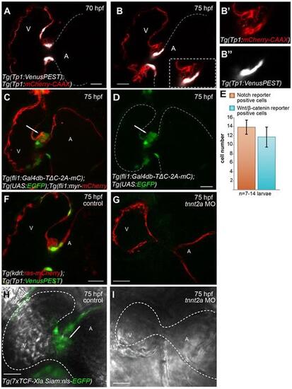
Fig. 5
- ID
- ZDB-FIG-231215-69
- Publication
- Pestel et al., 2016 - Real-time 3D visualization of cellular rearrangements during cardiac valve formation
- Other Figures
- All Figure Page
- Back to All Figure Page
|
Notch and Wnt/?-catenin signaling reporters exhibit different expression patterns in the forming cardiac valve and their activity in endocardial cells is dependent on cardiac contraction and/or blood flow. Sagittal planes of Tg(Tp1:VenusPEST);Tg(Tp1:mCherry-CAAX) hearts at 70 (A) and 75 (B) hpf. A magnification of the forming valve at 75 hpf is shown in B, inset; single channels are shown in B? and B?. The expression of both VenusPEST and mCherry-CAAX is driven by the Notch signaling-responsive element Tp1. At 75 hpf, in contrast to the mCherry-CAAX signal, which can be detected in both the ventricle and AVC (A,B,B?), the VenusPEST signal is only detectable in a subset of AVC cells lining the lumen (B,B?). At 70 hpf, the VenusPEST signal could also be detected in cells located at the border between the ventricle and AVC (A). (C,D) Sagittal plane of a 75 hpf Tg(fli1:Gal4db-T?C-2A-mC);Tg(UAS:EGFP);Tg(fli1:Myr-mCherry) heart. In contrast to the Notch reporter, the Wnt/?-catenin reporter is mainly expressed on the abluminal side of the valve (arrows). (E) Quantification of the number of AVC endocardial cells expressing VenusPEST of the Notch reporter or EGFP of the Wnt/?-catenin reporter. Data are mean▒s.d. of 14▒2 VenusPEST+ cells (n=7 larvae) and 12▒2 Wnt/?-catenin+ cells (n=14 larvae). (F-I) 75 hpf tnnt2a morphant hearts (G,I) and hearts of control injected larvae (F,H). Sagittal plane of Tg(kdrl:ras-mCherry);Tg(Tp1:VenusPEST) tnnt2a morphant heart shows that Tp1:VenusPEST+ cells observed in controls (F) are not detectable in non-beating hearts (G). Similarly, the TCF:EGFP signal in endocardial cells located at the AVC of control hearts (H, arrow) is downregulated in Tg(7xTCF-Xla.Siam:EGFP) tnnt2a morphants (I). A, atrium; V, ventricle. Scale bars: 20 Ám. |

