Fig. 1
- ID
- ZDB-FIG-231215-16
- Publication
- Edwards et al., 2023 - Hemato-vascular specification requires arnt1 and arnt2 genes in zebrafish embryos
- Other Figures
- All Figure Page
- Back to All Figure Page
|
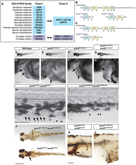
arnt1/2 double mutant embryos, but not arnt1 or arnt2 single mutants, lack circulating blood, have cardiac edema and atrial enlargement. (A) bHLH-PAS transcription factors act as heterodimers: a Class I protein and Class II protein interact and bind DNA. Asterisks indicate proteins encoded by genes present in zebrafish but absent in humans. (B,C) Generation of zebrafish with mutations in arnt1 and arnt2 genes. The amino acid sequences for the zebrafish Arnt1 (B) and Arnt2 (C) proteins are represented by the black line. Basic helix-loop-helix (bHLH), Per-Arnt-Sim (PAS) domains and the motif C-terminally located to the PAS (PAC) are represented by the labeled bubbles. The numbers below each domain indicate the starting and ending amino acid (AA) for each motif. (B) The arnt1bcm1 mutation causes a premature stop codon to occur at AA139 and the loss of the PAS-B, PAC and the majority of the PAS-A. The arnt1bcm2 mutation causes a premature stop codon to occur at AA250 and loss of the PAS-B and PAC domains. (C) The arnt2bcm3 mutation causes a premature stop codon to occur at AA287 and loss of the PAS-B and PAC domains. (D-I) Live images of embryos from the indicated genotype at 2 days post fertilization (dpf). All double homozygous arnt1bcm2/bcm2; arnt2bcm3/bcm3 embryos showed cardiac edema and atrial enlargement (G,G′). The wild-type (D,D′), arnt1bcm2/bcm2 (E,E′) and arnt2bcm3/bcm3 (F,F′) siblings of the double homozygous mutants (arnt1bcm2/bcm2; arnt2bcm3/bcm3) showed normal heart development. Black arrows in D′-G′ indicate the atrium and ventricle of the heart. Images are representative of seven different clutches, each clutch contained 20-300 embryos. All siblings of double homozygous mutants (arnt1bcm2/bcm2; arnt2bcm3/bcm3) had circulating blood (H; Movies 1, 2, 5 and 6) while arnt1/2 mutants lacked visible circulating blood cells (I; Movies 3, 4, 7 and 8). The black arrows in H indicate blood cells. The arrowheads in H and I indicate non-blood cells found in all embryos. (J-L) O-dianisidine staining to label blood cells in embryos 3 days post-fertilization shows that arnt1bcm2/bcm2; arnt2bcm3/bcm3 double homozygous mutants lack blood cells, while all other genotypes have clearly labeled blood cells. The arrows in J and K indicate the duct of Cuvier; the arrowheads in K indicate circulating blood cells. All embryos were derived from arnt1bcm2/+;arnt2bcm3/+ parents; images are representative of one clutch, containing 98 embryos. (J) n=96 of 98 embryos showed clear o-dianisidine staining and none of these embryos was an arnt1/2 mutant. (K) n=2 of 98 embryos showed a lack of o-dianisidine staining and both embryos were confirmed arnt1/2 mutants. For all images, anterior is towards the left and dorsal is towards the top, except for J, which shows a ventral view of the embryos. Scale bars: 1 mm in D-G; 100 μm in D′-G′ and H-L. |

