Fig. 3
- ID
- ZDB-FIG-231215-151
- Publication
- Demy et al., 2022 - Trim33 conditions the lifespan of primitive macrophages and onset of definitive macrophage production
- Other Figures
- All Figure Page
- Back to All Figure Page
|
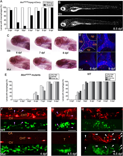
The production of macrophages from definitive hematopoiesis is delayed in moonshine mutants. (A) In vivo quantification of the percentage of sibling and monNQ039 mutant larvae with macrophages from 3 to 9 dpf, using a Tg(mpeg1:mCherry-F) background; n=84 sibling and 24 mutant larvae. (B) mCherry+ macrophages in live 8.5 dpf Tg(mfap4:mCherry-F) control sibling (top) and monTB222 mutant (bottom) larvae; maximum projection. (C) In situ hybridization for whole-mount detection of Csfr1a-expressing cells in control siblings (top) and monNQ039 mutants (bottom) at 6, 7 and 8 dpf. (D) Fluorescent immunodetection of L-plastin+ (leucocytes, red channel) macrophages in the brain of control siblings (top) and monNQ039 mutants (bottom) at 6 (left panels) and 9 (right panels) dpf; maximum projection, dorsal view. Nuclei are stained with Hoechst (blue channel). (E) In vivo quantification from 3 dpf to 9 dpf of the percentage of monNQ039 mutants (left graph) or wild-type controls (right graph) with mCherry+ macrophages under different experimental conditions aimed at removing primitive macrophages: injection of Pu1 MO, Irf8 MO or clodronate liposomes (Clod. Lip.), and metronidazole (Mtz)-mediated ablation in the Tg(mpeg1:Gal4;UAS:NfsB-mCherry) line; n=21 mutant and 88 wild-type larvae. (F-H) Lateral view of the (F) tail region or (G,H) thymus region of wild-type control (top) and mon mutants (bottom) expressing gata2b:Gal4;UAS:LifeAct-eGFP (hemogenic/HSPC-derived cells, green channel) and mpeg1:mCherry-F (macrophages, red channel) transgenes at 4 dpf (F), 4.5 dpf (G) and 6.5 dpf (H); maximum projection. Arrowheads indicate double-positive macrophages in controls (purple) and mutants (white); asterisks indicate pigment cells. Sib, sibling; Mut, mutant; hb, hindbrain; mb, midbrain; np, neuropil; CA, caudal artery; CHT, caudal hematopoietic tissue; CV, caudal vein; T, thymus. Scale bars: 50 Ám. |

