FIGURE

Figure 5

ID
ZDB-FIG-231204-40
Publication
Serrano et al., 2023 - Genetic model of UBA5 deficiency highlights the involvement of both peripheral and central nervous systems and identifies widespread mitochondrial abnormalities
Other Figures
All Figure Page
Back to All Figure Page
Figure 5

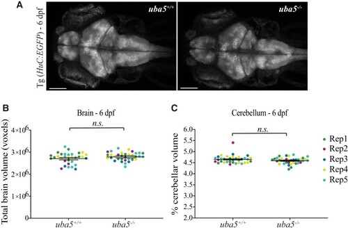
Quantification of total brain and cerebellar volume in uba5ex1s/ex1s animals at 6 dpf. (A) Maximum intensity projections of whole brain from Tg (Huc:EGFP); uba5+/+ and Tg (Huc:EGFP); uba5ex1s/ex1s 6 dpf zebrafish. (B) Total brain and (C) cerebellar volume analyses showed no significant changes in uba5ex1s/ex1s (brain volume n = 9, 4, 4, 8, 9; cerebellar volume n = 10, 4, 4, 8, 9) compared to uba5+/+ (brain volume n = 9, 3, 3, 7, 10; cerebellar volume n = 8, 4, 3, 9, 10). Each dot represents an individual zebrafish. Data represents raw data for five biological replicates plotted with the mean ± SD predicted by the mixed model statistical test. Scale bar = 100 µm. n.s., non-significant; Rep, replicate.

Expression Data
Gene:
Fish:
Anatomical Terms:
Stage: Day 6

Expression Detail
Antibody Labeling
Phenotype Data
Fish:
Observed In:
Stage: Day 6

Phenotype Detail
Acknowledgments
This image is the copyrighted work of the attributed author or publisher, and ZFIN has permission only to display this image to its users. Additional permissions should be obtained from the applicable author or publisher of the image. Full text @ Brain Commun