
Fig. 5
- ID
- ZDB-FIG-230831-17
- Publication
- Jackson et al., 2023 - Clinical, genetic, epidemiologic, evolutionary, and functional delineation of TSPEAR-related autosomal recessive ectodermal dysplasia 14
- Other Figures
- All Figure Page
- Back to All Figure Page
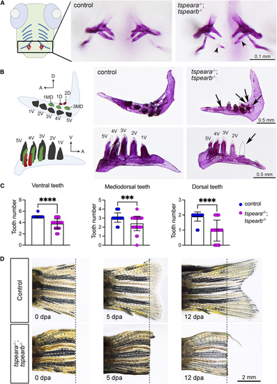
Zebrafish tspear double-knockout model (A) A schematic figure showing ventral view of larval zebrafish with ventral cartilages (blue) and teeth (red). Alizarin red S staining of pharyngeal teeth shows that a double homozygous mutant (tspeara?/?;tspearb?/?) displayed thinner and aberrant mineralized teeth (arrowhead) at 12 days post-fertilization (dpf). (B) Schematic figure for pharyngeal dentition of adult zebrafish shown in ventral view (top panel) and lateral view (bottom panel). Arrow indicates missing tooth. A, anterior; D, dorsal; MD, mediodorsal; V, ventral. (C) Quantification of the number of teeth in the ventral, mediodorsal, and dorsal rows. Each dot represents one animal. Control = 30 animals, mutant = 32 animals. Error bars: mean � SD. Difference was tested using two-tailed unpaired t test with Mann-Whitney test. ???p < 0.001, ????p < 0.0001. (D) Adult tail fins were amputated from wild-type control (n = 4) and double homozygous mutants (n = 4). Live fins were imaged at 0, 5, and 12 days post-amputation (dpa). Dashed line indicates the position of amputation. |
| Fish: | |
|---|---|
| Condition: | |
| Observed In: | |
| Stage Range: | Days 7-13 to Adult |