
Fig. 7
- ID
- ZDB-FIG-230430-89
- Publication
- Saelens et al., 2022 - An ancestral mycobacterial effector promotes dissemination of infection
- Other Figures
- All Figure Page
- Back to All Figure Page
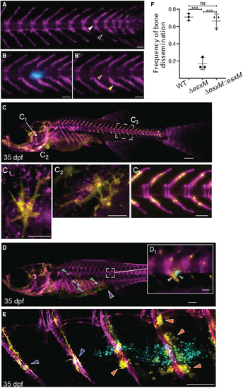
Ancestral esxM promotes spinal dissemination
(A) Representative image of sham-injected osteoblast line 3 weeks post-injection. Intervertebral spacing is tightly delineated (white arrowhead), and osteoblast signal at hemal arches is typical of healthy zebrafish (black arrowhead). Scale bar, 500 ?m. (B) Representative image of cerulean M. marinum-infected osteoblast line, three weeks post-infection (wpi) showing bone-associated infection. Scale bar, 100 ?m. (B?) Same animal as in (B), showing only osteoblasts. M. marinum has invaded into the intervertebral space, giving rise to a vertebral lesion (red arrowhead) and proliferation of osteoblasts proximal to infection (yellow arrowhead). Scale bar, 100 ?m. (C) Dual transgenic line at 35 dpf labeling osteoblasts in purple from the Sp7/osterix promoter and osteoclasts from the acp5a/TRAP promoter in yellow. Scale bar, 500 ?m. (C1-C3) Insets show tight association of osteoblasts and osteoclasts along the spine (C3). Yellow multinucleate osteoclasts are also visible in the head (C1 and C2). C1 inset scale bar, 500 ?m C2 inset scale bar, 50 ?. C3 inset scale bar, 100 ?m. (D) Intraperitoneal infection of juvenile animals with cerulean-fluorescent M. marinum results in frequent dissemination to spine (approximately 70% of animals). Scale bar, 500 ?m. (D1) High magnification view of boxed area of (D) showing association of granuloma with spine and alterations in normal osteoclast distribution. Scale bar, 100 ?m. (E) PACT-based clearing techniques in adults and juveniles enable high-resolution imaging of sites of bone disease. Confocal image of dorsal region of spine showing unaffected vertebrae with typical osteoclast behavior (dark blue arrowheads) and altered osteoclast behavior (orange arrowheads) in segments associated with granulomas (bacteria in cyan). Scale bar, 100 ?m. (F) Quantitation of dissemination to spine at 14 dpi in intraperitoneal infections. n ? 7 per group for 3 biological replicates. One-way ANOVA with Tukey?s post-test, data from three biological replicates, mean � SD shown. ***p < 0.001. |
| Genes: | |
|---|---|
| Fish: | |
| Condition: | |
| Anatomical Terms: | |
| Stage: | Days 30-44 |
| Fish: | |
|---|---|
| Condition: | |
| Observed In: | |
| Stage: | Days 30-44 |
Reprinted from Cell, 185(24), Saelens, J.W., Sweeney, M.I., Viswanathan, G., Xet-Mull, A.M., Jurcic Smith, K.L., Sisk, D.M., Hu, D.D., Cronin, R.M., Hughes, E.J., Brewer, W.J., Coers, J., Champion, M.M., Champion, P.A., Lowe, C.B., Smith, C.M., Lee, S., Stout, J.E., Tobin, D.M., An ancestral mycobacterial effector promotes dissemination of infection, 4507-4525.e18, Copyright (2022) with permission from Elsevier. Full text @ Cell