Figure 2.
- ID
- ZDB-FIG-230131-6
- Publication
- Brewer et al., 2022 - Macrophage NFATC2 mediates angiogenic signaling during mycobacterial infection
- Other Figures
- All Figure Page
- Back to All Figure Page
|
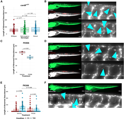
Pharmacological blockade of NFAT signaling inhibits angiogenesis while genetic ablation of card9 does not (A) Quantitation of angiogenic vessels along the trunk of card9 mutant, heterozygous, and wild-type animals 4 days post infection (dpi) with Mm-tdTomato. Each data point represents a single larva, n = 44 wild-type, 89 heterozygotes, 45 homozygous mutants. No statistically significant differences observed across the groups by Dunn?s Kruskal-Wallis multiple comparisons test with Holm error correction. Representative of three independent experiments. Additional independent replicates provided in Figures S1B and S1C. (B) Representative images from card9 wild-type and homozygous mutant zebrafish. Note the emergence of non-stereotypical vessels into the somites near the site of infection (inset). Arrowheads indicate regions of neovascularization. Scale bar, 250 ?m. (C) Quantitation of angiogenesis along the trunk of Mm-tdTomato-infected wild-type kdrl:eGFP zebrafish treated with either 125 nM FK506 or 0.0125% DMSO (vehicle) diluted in E3 medium. Each point represents mean vessel length within an independent biological replicate of 60?96 animals, with equal numbers of animals per replicate across three replicates. Box-and-whisker plot shows interquartile ranges. Statistical significance determined by Student?s t test. (D) Representative images of FK506- or vehicle-treated larvae. A single dose at a concentration that showed no developmental toxicity was provided immediately after infection. Doses at or above ?500 nM proved developmentally toxic. Arrowheads indicate ectopic vessels at the site of infection. (E) Quantitation of angiogenesis from wild-type kdrl:eGFP larval zebrafish injected with 2 mg/mL TDM in ?10?20 nL bolus or comparable volume of IFA vehicle alone. Treatment with 125 nM FK506 or 0.0125% ethanol (vehicle) for 2 dpi. Points shown are a random subset of 200 fish (50 fish per group) out of a total of 524 represented larvae to minimize data crowding. Each point represents a single larva with data pooled from three independent experiments. Statistics from Dunn?s Kruskal-Wallis multiple comparisons test with Holm error correction on the whole dataset. (F) Representative images from FK506-treated or ethanol (vehicle)-treated larvae at 2 dpi of TDM-injected larvae. FK506-treated larvae demonstrate a reduction in the degree of angiogenesis compared with the ethanol-treated group. Arrowheads indicate regions of angiogenesis. |
| Fish: | |
|---|---|
| Conditions: | |
| Observed In: | |
| Stage: | Day 5 |

