
FIGURE 4
- ID
- ZDB-FIG-221227-10
- Publication
- Palma et al., 2022 - Ontogenesis of the asymmetric parapineal organ in the zebrafish epithalamus
- Other Figures
- All Figure Page
- Back to All Figure Page
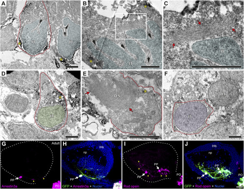
Parapineal cells show ultrastructural and immunohistochemical features distinctive from other epithalamic cells and suggestive of a photosensitive function in the adult zebrafish. (A?F) TEM images showing the cell types found in the adult (1.5?2 years old) zebrafish epithalamus: parapinealocytes ((A?C); nucleus labelled in light blue), pinealocytes ((D,E); nucleus labelled in green) and habenular cells ((F); nucleus labelled in pink). GFP-immunopositive parapinealocytes and pinealocytres display black DAB precipitates (yellow asterisks). The cell membrane is depicted as a red line. Parapinealocytes show a pear-shape cell body (red outline in (A)), a nucleus with indentations or clefts (black arrows in (A,B)), and a lamellar structure near the nucleus (red arrows in (C), which correspond to a high magnification view of the white rectangle depicted in (B)). Pinealocytes also show a pear-shape cell body (red outline in (D)) but the nucleus lacks indentations and shows an external segment characteristic of photoreceptors (red arrows in (E)). The cell body of habenular neurons is mostly occupied by its round and regular nucleus (E). (G?J) Immunofluorescence against Arrestin3a (G,H) and Rod opsin (I,J) in adult (1.5?2 years old) Tg(foxd3::GFP) zebrafish. Images correspond to dorsal views of confocal z-stack maximum projections, with anterior to the top, showing in the left epithalamus the fluorescence signal corresponding to Arrestin3a (magenta in (G)) and Rod opsin (magenta in (I)), or the merge fluorescence signals that also include the GFP of the pineal complex (green) and the DAPI/Hoechst nuclear staining that provides the left habenula tissue context (blue) (H,J). White arrows and yellow arrowheads indicate immunoreactive parapineal cell bodies and projections, respectively. Abbreviations: n (nucleus), os (outer segment). PO (pineal organ at the level of the stalk), PP (parapineal). Adult samples: Arrestin3a (n = 4), Rod opsin (n = 6). Scale bars, 1 �m (B, C), 2 �m (A,D?F) and 10 �m (G?J). |