|
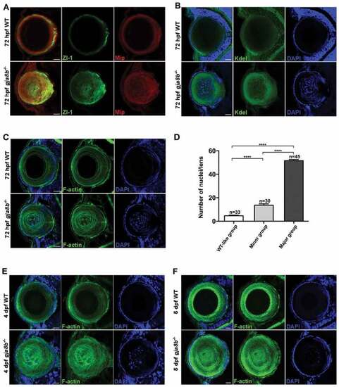
Ablation of Gja8b induces severe defects in organelle degradation of lens fiber cells. (A-C) Immunohistochemistry results showed that lens primary fiber cell markers (A, labeled by Zl-1 and Mip staining), endoplasmic reticulum (B, labeled by Kdel staining), cytoskeleton (C, labeled by F-actin staining), and nuclei (C, labeled by DAPI staining) remained in lens second fiber cells in gja8b mutants at 72 hpf. (D) The quantitative analysis of the average number of lens fiber cell nuclei shown in different gja8b mutants groups (WT-like group, minor group and major group) (mean � SEM, n ? 30 zebrafish for each group, ****p < 0.0001). (E and F) Immunohistochemistry results showed that cytoskeleton (labeled by F-actin staining), and nuclei (labeled by DAPI staining) remained in the center of lens in gja8b mutants at 4 dpf (E) and 5 dpf (F). Scale bars: 50 ?m
|