
Figure 2
- ID
- ZDB-FIG-221214-180
- Publication
- Burgoyne et al., 2022 - Changes in Mitochondrial Size and Morphology in the RPE and Photoreceptors of the Developing and Ageing Zebrafish
- Other Figures
- All Figure Page
- Back to All Figure Page
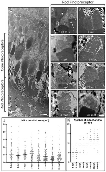
Rod inner segments of early embryonic zebrafish have compact mitochondria with a morphology different from other ages. (A) Electron microscopy image of 12 mpf retina between the RPE and ONL that includes rod and cone photoreceptor outer (POS) and inner segments. (B?I) Images of rod photoreceptor inner segments at 5 dpf through to 36 mpf. (B) At 5 dpf, the mitochondria are bundled together in a spherical like arrangement and there is a morphology change by (C) 8 dpf, with further changes by (D) 1 mpf as the mitochondria become less clumped together. There is (J) a reduction in rod inner segment mitochondrial size and (K) an increase in mitochondria number with time. Measurements were acquired from n = 3 zebrafish and included (J) >83 mitochondria and (K) n = 15 rods at each timepoint. (J,K) Statistical significance determined by one way-ANOVA (J,K) with p < 0.0001. Scale (A) 10 ?m, (B?I) 1 ?m. |