
FIGURE
Fig. 6
- ID
- ZDB-FIG-221018-222
- Publication
- Keil et al., 2021 - Heparan sulfate proteoglycan expression in the regenerating zebrafish fin
- Other Figures
- All Figure Page
- Back to All Figure Page
Fig. 6
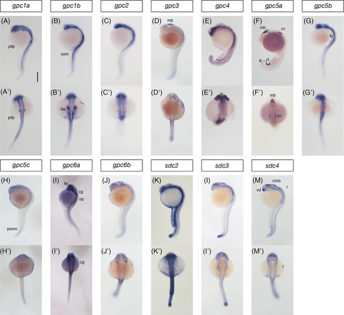
Prim-5 embryo staining controls for in situ probes. 24 hpf embryos are shown in lateral (A-M) and dorsal (A?-M?) views. Embryos in E/E? and F/F? are slightly younger. Scale bar 250 ?m. pllp, posterior lateral line primordium; som, somites; ba, branchial arch; mb, midbrain; ov, otic vesicle; cl, cloaca; ff, fin fold; fp, floor plate; psom, posterior somites; te, telencephalon; cg, cranial ganglion; vd, ventral diencephalon; mhb, midbrain hindbrain boundary; r, rhombomeres
|
Expression Data
Expression Detail
Antibody Labeling
Phenotype Data
Phenotype Detail
Acknowledgments
This image is the copyrighted work of the attributed author or publisher, and
ZFIN has permission only to display this image to its users.
Additional permissions should be obtained from the applicable author or publisher of the image.
Full text @ Dev. Dyn.