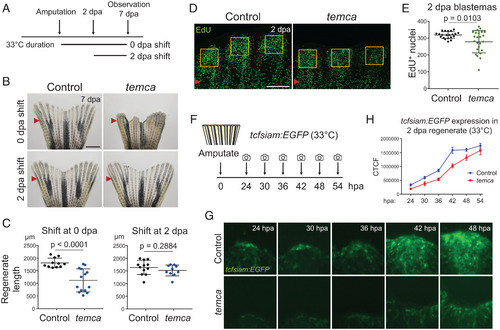
Early fin regeneration is impaired in temca mutants. (A) Schematic of temperature shift assay. (B) Representative whole-mount images of control (heterozygotes) and temca fin regenerates at 7 dpa shifted to 33 °C right after amputation (0 dpa shift) or at 2 dpa (2 dpa shift). (C) Quantification of fin regenerate lengths at 7 dpa (n = 12, 14, 12, and 11 for 0 dpa shift control and temca and for 2-dpa shift control and temca, respectively). (D) Representative EdU-staining images of control and temca fin regenerates at 2 dpa. (E) Quantification of EdU+ cells in blastema area of 2 dpa fin regenerates (n = 8). (F) Schematic showing amputation and imaging time points of tcfsiam:EGFP transgenic reporter line. (G) Representative whole-mount images of tcfsiam:EGFP control (heterozygotes) and temca fin regenerates at 24, 30, 36, 42, and 48 h postamputation (hpa) at 33 °C. (H) Quantification of tcfsiam:EGFP signal intensity (n = 10 and 9 for control and temca, respectively). Red arrowheads indicate amputation planes. Scale bars, 1 mm in B; 150 µm in D. Data are presented as mean ± SD. Student’s unpaired two-tailed t test.
|
