
Fig. 2
- ID
- ZDB-FIG-220520-48
- Publication
- Brunsdon et al., 2022 - Aldh2 is a lineage-specific metabolic gatekeeper in melanocyte stem cells
- Other Figures
- All Figure Page
- Back to All Figure Page
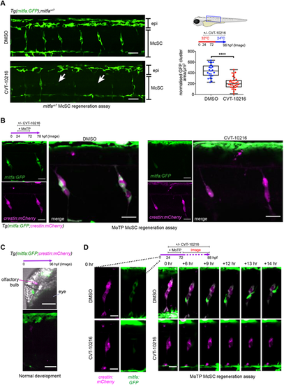
Live imaging captures the McSC requirement for Aldh2 to generate progeny. (A) An ALDH2 inhibitor (CVT-10216) causes loss of mitfa:GFP expression in McSCs, while dorsal stripe epithelial (epi) GFP+ melanoblasts remain. Representative confocal stack images of McSCs at the niche after 24 h regeneration with or without CVT-10216 treatment. The average mitfa:GFP niche area μm2/somite was quantified per embryo (one data point) Boxes indicate median and quartiles; whiskers span minimum to maximum values. McSCs with very low to no GFP signal are indicated with arrows. Scale bars: 50 μm, three experimental replicates.****P<0.0001 (unpaired, two-tailed t-test). (B) McSCs maintain neural crest identity when treated with an ALDH2 inhibitor (CVT-10216). Confocal stack images of McSC niches in CVT-10216-treated Tg(mitfa:GFP;crestin:mCherry) embryos after 6 h washout of MoTP. Two experimental replicates, five or more embryos used per condition, representative images shown. Scale bars: 50 µm. (C) 96 hpf non-regenerating Tg(mitfa:GFP;crestin:mCherry) embryos (same age as B) still express crestin:mCherry in the olfactory bulb and mitfa:GFP in embryonic epithelial melanoblasts [labelled in head (top) and trunk (bottom)], but no longer express these transgenes in McSC niches. Representative images of three embryos are shown. Scale bars: 50 µm. (D) Time-lapse stills of individual regenerating McSCs at the niches. Tg(mitfa:GFP; crestin:mCherry) embryos with or without CVT-1016 were imaged from 2 h post-MoTP washout. In a control embryo, an McSC undergoes cell division and a new mitfa:GFP-high cell migrates upwards towards the epidermis (see Movie 3). In a CVT-10216-treated embryo, mitfa:GFP expression is absent and migration is not observed (see Movie 4). Scale bars: 20 µm. |