|
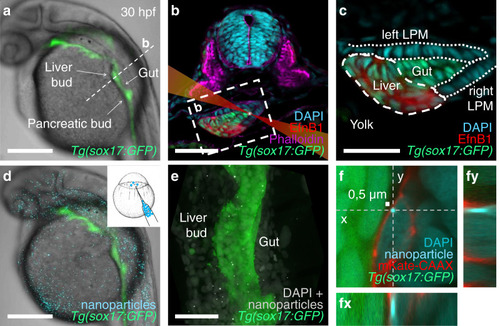
Injected nanoparticles distribute throughout the embryo, including the foregut region in 28–30 hpf zebrafish embryos.a Transgenic tg(-0.5 sox17:GFP)zf99 visualizes the endoderm in a living zebrafish embryo, including the foregut organ primordia. b Transverse section through the foregut region (dashed line in a) with schematized laser beam path (red) and bead (white). The foregut endoderm expresses transgenic sox17:GFP (green) and liver progenitors EphrinB1 (red); DAPI marks nuclei (cyan); and Phalloidin marks F-actin (magenta). c Magnification of the foregut region shown in (b) (white dashed rectangle). d 0.5 µm polystyrene particles (cyan) microinjected at the one-cell stage distribute throughout the embryo, including the endoderm (green), without causing apparent morphological defects. e Maximum intensity projection of confocal z-stack showing nanoparticle distribution (white) in the foregut region, GFP marks the endoderm. f Confocal image of a representative nanoparticle (light blue) located in the cytoplasm between the nucleus (darker blue) and plasma membrane (red) in a fixed embryo. (fy) and (fx) show orthogonal views in the “yz” plane (dashed line “y” in (f)) and “xz” plane (dashed line “x” in (f)), respectively. Scale bars a, d: 250 µm; b, e: 50 µm; c: 30 µm; f: 0,5 µm (scale bar next to bead).
|