|
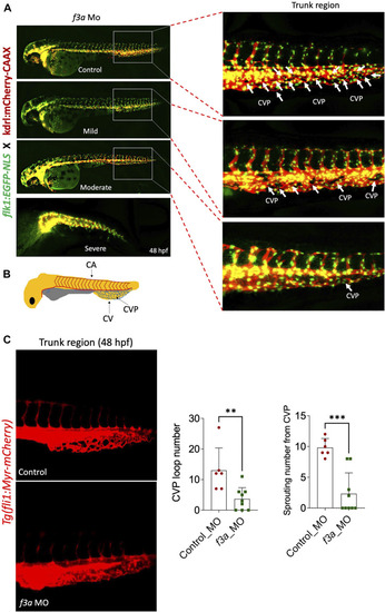
f3a knockdown shows delayed angiogenesis: To study vascular development, endothelial-specific transgenic lines of zebrafish (flk1:egfp-NLS/kdrl:mCherry-CAAX) embryo were used to knockdown f3a. (A) Images represent MO-mediated knockdown of f3a expression resulting in morphological abnormalities. MO phenotypes: control MO injected (control MO), and mild, moderate, and severe phenotypes. Differences in caudal vein plexus (CVP) in the trunk regions are indicated by white arrows. (B) Schematic diagram depicts to identify Caudal vein (CV), caudal vein plexus (CVP) and Central aorta (CA). (C)Tg(fli1:Myr-mCherry) line was used to quantify CVP loop number and Sprouting number from the CVP at 48 hpf. Embryos were randomly picked from ∼60 MO-injected (6 from control and 9 from f3a MO) embryos from control and f3a.
|