|
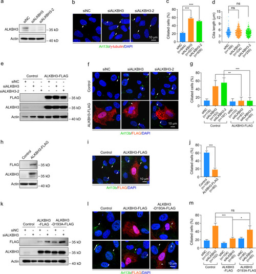
ALKBH3 inhibits ciliogenesis through its m<sup>1</sup>A demethylation activity.a–d RPE-1 cells were transfected with control or ALKBH3 siRNAs for 48 h in DMEM/F12 medium with 10% serum (normal conditions) and then subjected to western analysis or immunofluorescence. Western blotting of ALKBH3 protein (a). Representative images of RPE-1 cells with anti-Arl13b (green) and γ-tubulin (red) antibodies (b). Quantification analysis of the percentage of ciliated cells (c). Cilia length was determined by Image J software (d). e–g, k–m RPE-1 cells treated with the indicated siRNAs for 24 h were transfected with control, ALKBH3-FLAG, or ALKBH3-D193A-FLAG plasmid for another 24 h in normal conditions, and then subjected to western analysis or immunofluorescence. h–j RPE-1 cells transfected with the indicated plasmids for 24 h in normal conditions were treated with serum starvation for another 24 h and then applied for western analysis or immunofluorescence. Western blotting of FLAG and ALKBH3 protein was presented (e, h, k). Representative confocal images of RPE-1 cells with anti-Arl13b (green) and anti-FLAG (red) antibodies were shown (f, i, l). The percentage of ciliated cells in control or FLAG-positive group was analyzed (g, j, m). Actin was served as a loading control. DNA was stained by DAPI (blue). Cilia are indicated by white arrows. Scale bars, 10 μm. n, the number of total cells calculated. Data are presented as the means ± SD from at least three independent experiments. Student’s t-test; ns not significant; *P < 0.05, **P < 0.01, ***P < 0.001.
|