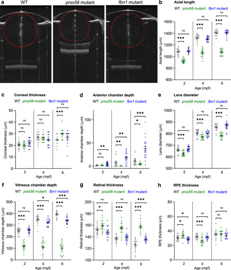
Loss of prss56 or fbn1 leads to distinctive biometrical changes in the eye. SD-OCT recordings of size-matched 2 to 6 mpf zebrafish biometrics of ocular compartments. Individual metrics of each compartment of the eye were corrected for the tissue-specific refractive index. (a) A single B-scan image of a typical 4 mpf zebrafish eye of respectively a WT, prss56 mutant, and fbn1 mutant. (b) Eyes of the prss56 mutants were significantly reduced in axial length compared with WT eyes at 2 mpf (effect size = −157 µm; P < 0.001), 4 mpf (effect size = −260 µm; P < .001), and 6 mpf (effect size = −330 µm; P < 0.001). The fbn1 mutant eyes showed no significant alteration relative to WT. (c) The corneal thickness of 6 mpf prss56 mutants was significantly increased (effect size = 5 µm; P < 0.001). No significant changes in corneal thickness were found in the fbn1 mutants. d The ACD in the prss56 mutants was significantly decreased at 4 mpf (Effect size = −7 µm; P < 0.01) and 6 mpf (Effect size = −6 µm; P < 0.05). The ACD was significantly increased in the fbn1 mutants at 2 mpf (effect size = 3 µm; P < 0.05), 4 mpf (effect size = 11 µm; P < 0.01), and 6 mpf (effect size = 27 µm; P < 0.001). (e) The lens diameter was significantly reduced in the prss56 mutants at 2 mpf (Effect size = −32 µm; P < 0.05), 4 mpf (effect size = −34 µm; P < 0.01), and 6 mpf (effect size = −73 µm; P < 0.001). The fbn1 mutant eyes showed no significant alteration in lens diameter relative to WT. f The VCD of the prss56 mutants was significantly reduced at 2 mpf (effect size = −132 µm; P < 0.001), 4 mpf (effect size = −237 µm; P < 0.001), and 6 mpf (effect size = −289 µm; P < 0.001). The fbn1 mutants showed a decrease in VCD at 6 mpf (effect size = −51 µm; P < 0.001). (g) The retinal thickness was larger in prss56 mutants at 2 mpf (effect size = 7 µm; P < 0.05), 4 mpf (effect size = 19 µm; P < 0.001), and 6 mpf (effect size = 32 µm; P < 0.001) and remained stable (range, 155–160 µm), whereas the WT retina is thinning over time (152 µm at 2 mpf; 137 µm at 4 mpf; 126 µm at 6 mpf). The retinal thickness was relatively increased in 6 mpf fbn1 mutants (effect size = 10 µm; P < 0.01). (h) The RPE thickness was significantly altered in 2 mpf (effect size = 3 µm; P < 0.05) and 4 mpf (effect size = −5 µm; P < 0.001) prss56 mutants while no significant differences were found in the fbn1 mutants. Sample size: n = 20 eyes for each genotype and time point. See Supplementary Table S2 for all statistics. Error bars: standard error of the mean. Significance: ns = not significant, *P < 0.05, **P < 0.01, ***P < 0.001. Scale bar, 100 µm. Mpf, months postfertilization; ACD, anterior chamber depth; VCD, vitreous chamber depth; RPE, retina pigmented epithelium.
|
