|
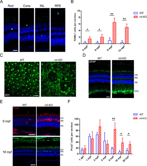
Progressive degeneration and regeneration in the <italic toggle='yes'>nrl</italic>-KO retinas.(A) The TUNEL assay results revealed that multiple types of retinal cells, including rods, cones, RPE, and inner retinal cells, undergo apoptosis in the nrl-KO retinas. Representative images are shown. Scale bar: 20 μm. (B) Quantitation of the apoptotic cells per section from 5 mpf to 13 mpf. The results are shown as mean with SD (n = 3). *, p < 0.05; **, p < 0.01. (C) RPE morphology in the WT and nrl-KO zebrafish at 20 mpf are shown, as assessed by immunostaining the retinal whole-mounts for ZO-1 Scale bar: 20 μm. (D) The up-regulation of GFAP in nrl-KO retinas as detected by immunostaining. CN, cone nuclear layer; RN, rod nuclear layer; INL, inner nuclear layer; GCL, ganglion cell layer. Scale bar: 25 μm. (E) Regeneration of photoreceptors was more active in the nrl-KO retinas at 9 mpf and 18 mpf, as reflected by the increase in the number of Pcna-positive cells (proliferating cells) in the ONL, compared with the WT levels. Scale bars: 50 μm. (F) Quantitation of the Pcna+ cells located in the ONL per section. The results are shown as mean with SD (n = 4) from 7 dpf to 29 mpf. *, p < 0.05; **, p < 0.01.
|