|
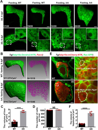
The lvh mutant exhibits hepatomegaly and enlarged hepatocyte size under fasting. (A) Confocal 3D projection and 2D single-optical section images of the liver under feeding and fasting in the wild-type or lvh mutant at 9 dpf. Higher magnification images showing single hepatocytes are displayed upright. (B) 3D reconstruction images showing the liver volumes and hepatocyte nuclei of the wild-type and lvh mutant at 9 dpf. (C) Unpaired Student?s t-test for the liver volume of wild-type (n = 5) and lvh (n = 5). (D) Unpaired Student?s t-test for the number of hepatocytes per liver of wild-type (n = 5) and lvh (n = 5). (E) Single-optical section images showing livers of the wild-type and lvh mutant at 9 dpf. Higher magnification images of single hepatocytes (dashed frames) are displayed. (F) Unpaired Student?s t-test for single hepatocyte volume in the wild-type (n = 5) and lvh (n = 5). NS, not significant. ****P<0.0001. WT, wild-type. Data are represented as mean�SD. Scale bars, 50 ?m.
|