|
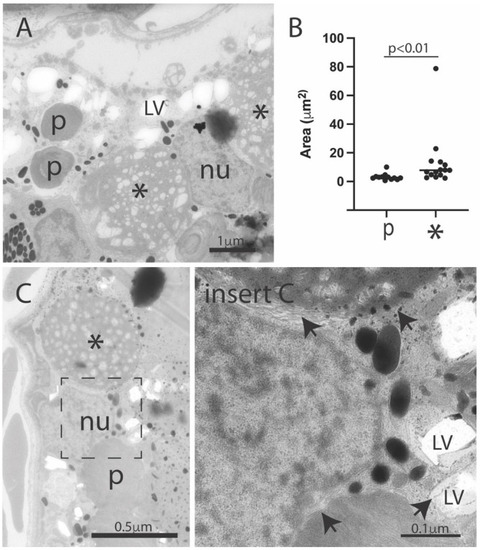
Ultrastructural features in the retinal pigment epithelium of the DJ-1-deficient retina. Electron micrographs of the RPE area of DJ-1 knockouts (A,C). Images show both phagosomes (p) and the larger electron-dense structures (*), in addition to numerous small electron lucent vacuoles (LV) in RPE. Phagosomes and electron lucent vacuoles were surrounded by membranes (arrows), but no clear limited membrane was observed limiting the electron-dense structures. (B) Quantitation of area of phagosomes (p) and electron-dense structures (*); p < 0.01. Mann-Whitney-test, n = 12 (WT) and 15 (KO_DJ-1); p, phagosome; * electron-dense structure; LV, electron-lucent vacuole; nu, RPE nucleus.
|