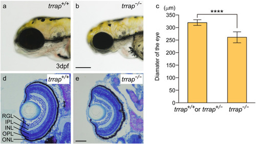
Small eyes in the trrap-zebrafish mutant. (a) Wild-type fish (trrap+/+) at 3 dpf. (b) trrap mutants (trrap−/−) at 3 dpf. Scale bar, 200 μm. (c) The eye diameters in larvae containing the wild-type allele (trrap+/+: n = 2; trrap+/−: n = 6) and larvae containing trrap mutant alleles (trrap−/−: n = 8) were measured. The error bars indicate the standard deviation. Asterisks indicate statistical significance between the wild-type and the mutant zebrafish. ****P < 0.0001. (d,e) Cross-sections of the wild-type (trrap+/+) (d) and the trrap-mutant (trrap−/−) zebrafish (e) at 3 dpf were stained with toluidine blue (0.1%). The eye diameter was reduced in the trrap mutants, whereas the laminated retinas consisting of three layers (RGL, INL and ONL) and two plexiform layers (IPL and OPL) developed normal in the wild-type and mutant zebrafish. RGL retinal ganglion cell layer, IPL inner plexiform layer, INL inner nuclear layer, OPL outer plexiform layer, ONL outer nuclear layer. Genomic DNA was isolated from individual caudal fins, and genotyping was performed by genomic PCR. Scale bar, 50 μm.
|
