
Figure 5
- ID
- ZDB-FIG-211224-43
- Publication
- Robey et al., 2021 - Characterization and tissue localization of zebrafish homologs of the human ABCB1 multidrug transporter
- Other Figures
- All Figure Page
- Back to All Figure Page
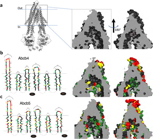
3D homology modeling of amino acid similarity in the binding pocket of zebrafish Abcb4 and Abcb5. (a) 3D modeling of the human P-gp structure that models P-gp in complex with taxol and the Fab of the UIC2 antibody (PDB:6QEX). The model is split into two halves to provide a clearer view. Amino acid differences in the transmembrane regions of the models of human P-gp and zebrafish Abcb4 (b) and Abcb5 (c) are shown. In (b) and (c), on the left side linear 2D models of Abcb4 and Abcb5 are shown. Amino acids (filled circles) similar to human P-gp are shown in green (most similar), yellow (fairly similar) or red (least similar). |