
FIGURE 2
- ID
- ZDB-FIG-211111-33
- Publication
- Camacho-Macorra et al., 2021 - Mosmo Is Required for Zebrafish Craniofacial Formation
- Other Figures
- All Figure Page
- Back to All Figure Page
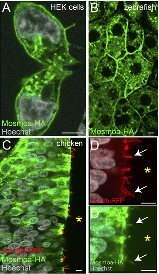
Figure 2. Mosmoa localizes at the plasma membrane, endosomes, and primary cilia. (A) Example of human embryonic kidney (HEK) cells transfected with mosmoa-HA, immunostained for ?-HA (green), and counterstained with Hoechst (white). (B) Dorsal view of a zebrafish gastrula (7 hpf) injected with mosmoa-HA mRNA and immunostained for ?-HA (green). (C?D?) Transversal sections of chick embryo neural tubes co-electroporated with mosmoa-HA and the cilia marker arl13b-RFP. In both HEK cells and zebrafish EVL cells Mosmoa-HA signal localizes at the plasma membrane and in endo-vesicles. In HH14 chick embryos, neural tube Mosmoa-HA is also observed in the in the Arl13b-positive cilia (white arrows) (D,D?). Yellow asterisk marks the neural tube ventricle (C?D?). Scale bars: 5 ?m. |