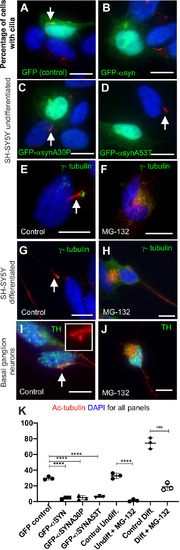
Aggresomes inhibit ciliogenesis. All panels: DNA/nuclei stained with DAPI (blue), green label is indicated in each panel, cilia (white arrows) can be identified by acetylated tubulin (red). (A–F) When transfected with a GFP-α-syn expression plasmid (α-syn and A30P and A53T familial mutants) or treated with MG132, cilia formation is inhibited in undifferentiated SH-SH5Y cells (B, C and D versus A, F versus E). In the presence of aggresomes differentiated SH-SY5Y cells are no longer able to form cilia (H versus G). When treated with MG132, TH-positive basal ganglion neurons are no longer able to form cilia (J versus I). The acetylated tubulin signal (red) in J and I is overexposed to ensure no cilia were missed. (K) Quantification of ciliation: GFP expressing versus GFP-α-syn expression, (P=0.0003, by one-way ANOVA, 100 cells, n=3); undifferentiated SH-SY5Y, untreated versus MG132, (P=0.0001, by Student's t-test, 100 cell, n=3); differentiated SH-SY5Y, untreated versus MG132, (P=0.0001 by Student's t-test, 100 cells counted, n=3). Scale bars: 10 μM.
|
