
FIGURE
Figure 2
- ID
- ZDB-FIG-210915-89
- Publication
- Schellens et al., 2021 - Zebrafish as a Model to Evaluate a CRISPR/Cas9-Based Exon Excision Approach as a Future Treatment Option for EYS-Associated Retinitis Pigmentosa
- Other Figures
- All Figure Page
- Back to All Figure Page
Figure 2
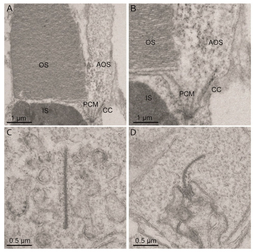
Eys localization in adult zebrafish photoreceptor cells. Immunoelectron microscopy images of adult zebrafish retinas show that Eys localizes at (A,B) the connecting cilium, periciliary membrane, cone accessory outer segments, and (C,D) photoreceptor ribbon synapses. CC: connecting cilium, PCM: periciliary membrane, AOS: accessory outer segment, IS: inner segment, OS: outer segment. |
Expression Data
| Gene: | |
|---|---|
| Antibody: | |
| Fish: | |
| Anatomical Terms: | |
| Stage: | Adult |
Expression Detail
Antibody Labeling
Phenotype Data
Phenotype Detail
Acknowledgments
This image is the copyrighted work of the attributed author or publisher, and
ZFIN has permission only to display this image to its users.
Additional permissions should be obtained from the applicable author or publisher of the image.
Full text @ Int. J. Mol. Sci.