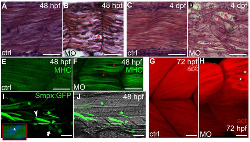
smpx knockdown disturbs the proper fiber arrangement during development. (A?D) side views, anterior left, dorsal top. Paraffin sections of Control-MO (A,C) and smpx-MO (B,D) injected embryos at 48 hpf (A,B) and 4 dpf (C,D). The red asterisks indicate alterations in the muscle array. (E?H) side views, anterior left, dorsal top. Confocal Z-stacks taken from whole-mount embryos (48 hpf) and larvae (72 hpf) labelled with (E,F) myosin heavy chain (MHC) antibody for slow fibers and (G,H) phalloidin (act) for actin filaments of the fast fibers. The red (F) and white (H) asterisks indicate alterations in the normal muscle array (controls in (E,G), respectively). (I,J) representative lateral view of an embryo injected with the construct encoding the Smpx:GFP chimera (see Figure S1A for construct details); muscle fibers (arrowhead) and epithelial cells (arrow) are both uniformly painted with no accumulation of Smpx:GFP in the nuclei. Inset: magnification of a slow fiber with the nucleus (asterisk) labelled with DAPI (blue). Anterior left, dorsal top. Scale bars = 25 ?m in (A?D); 20 ?m in (E,F); 20 ?m in (G,H); 100 ?m in (I,J).
|
