|
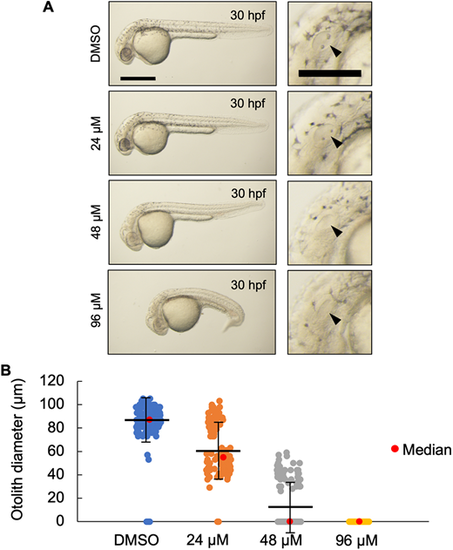
(A) Live imaging of 30?hpf wild-type embryos treated with the indicated dose of U18666A starting at 4?hpf. U18666A-treated embryos exhibited a dose-dependent phenotype ranging from smaller otoliths at 24??M to no otoliths, abnormal head, no circulating blood cells, and curved/twisted body axis at 96??M. Arrowheads indicate the otic vesicle. Scale bars: 500??m (whole embryo); 200??m (otic vesicle). (B) Measurement of otolith diameters from DMSO- and U18666A-treated wild-type embryos at 30?hpf. n=118 (DMSO), 118 (24??M), 114 (48??M) and 94 (96??M) (n=2 experiments). Two otoliths per embryo were measured. 0 indicates that otolith was not detectable. P<0.0001 for each treatment group compared with DMSO (Kruskal?Wallis test).
|