|
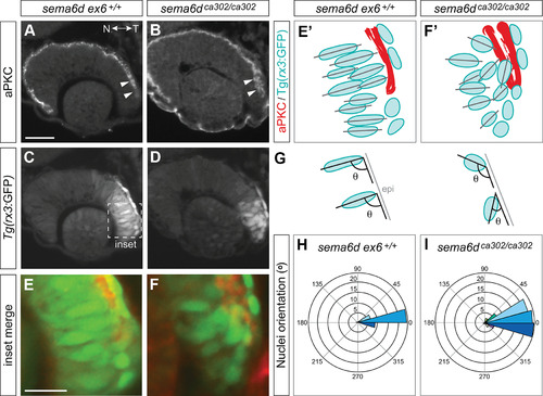
Temporal neural retina is disorganized in sema6d mutants. A–F, Immunohistochemistry for the apical marker aPKC (A, B) of wild-type and sema6d mutant (C, D) Tg(rx3:GFP) embryos. Magnified view of merge of labels in temporal eye (boxed area in C; E, F). E’–F’, Schematic of E, F showing ventricle as marked by aPKC immunoreactivity (red), labeled GFP (blue) cells, and their orientation (black lines). In sema6d mutants, there is a failure of the temporal ventricle to seal (arrowheads in B) and disorganization of temporal neural retina cells (D, F, F’). G, H, Distributions of the angles made by the long axis of DAPI-labeled nuclei with the basal temporal retinal neuroepithelium at 24 hpf. Average of the distributions in mutants (I, n = 12 embryos) and wild-type sibling (H, n = 13 embryos) are significantly different (p < 0.0001, χ2 contingency test). Circles indicate the average numbers of nuclei found in each 15° bin. Scale bars: 50 μm (A) and 20 μm (E). N: nasal, T: temporal.
|