
Fig. 3
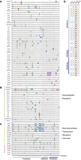
Figure 3. Neuron Subtype Diversity at 15 dpf (A?C) Violin plots of select marker gene expression in identified brain neuron subtypes at 15 dpf. Retinal neurons and nascent neurons are omitted from the analysis. Cluster numbers are indicated at the bottom along with their inferred spatial location in the brain. Cluster 76 has unknown spatial location. Detailed cluster descriptions are in Table S1 and can be explored interactively at https://github.com/brlauuu/zf_brain. (A) Expression of transcription factors. (B) Expression of neuropeptides and their receptors. (C) Expression of genes involved in neuron electrophysiology. (D) Matrix showing overlap of neuron subtypes identified at 15 dpf and earlier larval (5 and 8 dpf) or later juvenile (25 dpf [Raj et al., 2018b]) stages. The cluster number at 15 dpf is shown, and an orange circle indicates that the subtype is detected in another stage. |