
Fig. 9
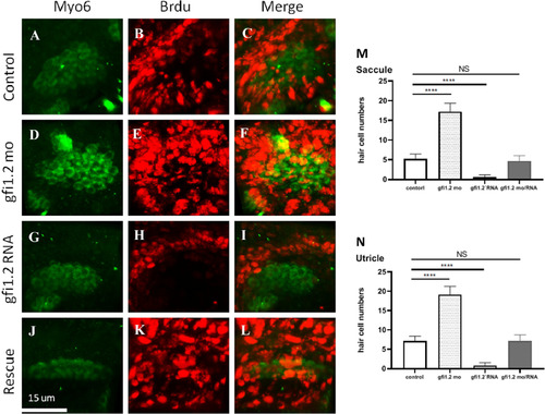
Myo6 (green) is the marker for hair cells, and BrdU positivity (red) represents proliferating cells. Merged images labeled proliferating hair cells in the specimen. The presence of proliferating hair cells in wild-type zebrafish (control group) was demonstrated in (A–C). With gfi1.2 MO treatment, the proliferating hair cells increased in number (D–F). On the other hand, gfi1.2 mRNA treatment suppressed the proliferation of hair cells (G–I). gfi1.2 mRNA-induced hair cell suppression was rescued by adding gfi1.2 MO (J–L). Proliferating hair cells in (M) saccule and (N) utricle are further quantified. (For interpretation of the references to color in this figure legend, the reader is referred to the web version of this article.) |
| Fish: | |
|---|---|
| Knockdown Reagents: | |
| Observed In: | |
| Stage: | Protruding-mouth |
Reprinted from Hearing Research, 396, Yu, R., Wang, P., Chen, X.W., The role of gfi1.2 in the development of zebrafish inner ear, 108055, Copyright (2020) with permission from Elsevier. Full text @ Hear. Res.