
FIGURE
Figure 2—figure supplement 2.
- ID
- ZDB-FIG-201021-7
- Publication
- Helker et al., 2020 - Apelin signaling drives vascular endothelial cells towards a pro-angiogenic state
- Other Figures
-
- Figure 1
- Figure 1—figure supplement 1.
- Figure 1—figure supplement 2.
- Figure 1—figure supplement 3.
- Figure 2
- Figure 2—figure supplement 1.
- Figure 2—figure supplement 2.
- Figure 2—figure supplement 3.
- Figure 3
- Figure 3—figure supplement 1.
- Figure 3—figure supplement 2.
- Figure 4
- Figure 4—figure supplement 1.
- Figure 5
- Figure 5—figure supplement 1.
- All Figure Page
- Back to All Figure Page
Figure 2—figure supplement 2.
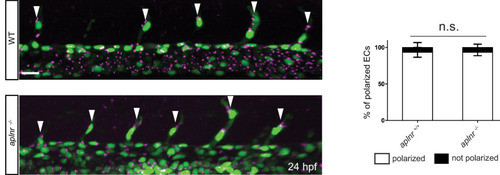
Confocal projection images of the trunk region of Tg(kdrl:NLS-EGFP); Tg(fli1a:B4GALT1-mCherry) embryos at 24 hpf. Apelin deficiency does not cause obvious defects in EC polarity (aplnr +/+, n = 6; aplnr -/-, n = 8). Arrowheads point to polarized tip cells in the ISVs. Scale bar: 20 �m.
|
Expression Data
Expression Detail
Antibody Labeling
Phenotype Data
Phenotype Detail
Acknowledgments
This image is the copyrighted work of the attributed author or publisher, and
ZFIN has permission only to display this image to its users.
Additional permissions should be obtained from the applicable author or publisher of the image.
Full text @ Elife