
Fig. 1
- ID
- ZDB-FIG-200818-7
- Publication
- Mushtaq et al., 2020 - Cell stemness is maintained upon concurrent expression of RB and the mitochondrial ribosomal protein S18-2
- Other Figures
- All Figure Page
- Back to All Figure Page
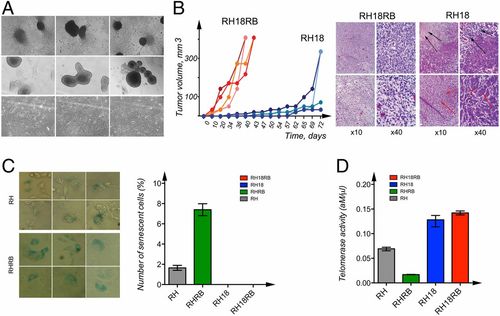
Characterization of cell lines generated from Rb1?/? MEFs. (A) Phase-contrast images of RH18RB cells in cell culture dishes (Upper) and in bacterial petri dishes (Middle). Phase-contrast images of RH18 cells in cell culture dishes (Lower). (B) The kinetics of tumor growth of RH18 and RH18RB cells as indicated in SCID mice (Left). Sections of the tumors produced by RH18 and RH18RB cells were stained with hematoxylin and eosin. The RH18RB cells generated aggressive fibrosarcomas. The RH18 tumors contained large necrotic areas (Right, black arrows). A proportion of the cells in these tumors resembled epithelium-like cells (Right, red arrows). (C) Staining with ?-galactosidase for the identification of senescent RHRB (Top Left) and RH (Bottom Left) cultures. Arrows indicate cells producing ?-galactosidase. The number of senescent cells was analyzed using column analysis and an unpaired t test (Right). (D) The telomerase activity in RH, RHRB, RH18, and RH18RB cells was determined via the QTD method using qPCR. Each bar represents the mean telomerase activity of each cell culture. Reactions were performed twice, with each cell line assessed in triplicate. Statistical analysis was performed as in C. |