FIGURE

Fig. 3

ID
ZDB-FIG-200629-20
Publication
Chen et al., 2020 - Capn3 depletion causes Chk1 and Wee1 accumulation and disrupts synchronization of cell cycle reentry during liver regeneration after partial hepatectomy
Other Figures
All Figure Page
Back to All Figure Page
Fig. 3

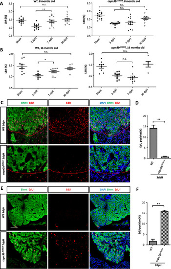
Delayed hepatocyte proliferation in capn3bΔ19Δ14 at the resection site after PH. a, b Comparison of LBRs at 3-, 7- and 30-dpH in WT and capn3bΔ19Δ14 fish operated at the age of 8-months-old (a) or16-months-old (b). Sham control: cut through the abdominal skin only. c-f Co-staining of EdU and Bhmt (liver marker) at the wounding site of the liver in WT and capn3bΔ19Δ14 fish at 3dpH (c) and 7dpH (e). Red dashed line: outlining the cutting site. DAPI: staining nuclei. Scale bar: 50 μm. Corresponding statistical data of the EdU-positive cells were provided (d, 3dpH; f, 7dpH). EdU was injected 24 h before harvesting at 3dpH or 7dpH. Three sections per fish and total three fish were evaluated for each genotype in each case. Student’s T-test for statistical analyses, *, p < 0.05; **, p < 0.01

Expression Data

Expression Detail
Antibody Labeling
Phenotype Data
Fish:
Condition:
Observed In:
Stage: Adult

Phenotype Detail
Acknowledgments
This image is the copyrighted work of the attributed author or publisher, and ZFIN has permission only to display this image to its users. Additional permissions should be obtained from the applicable author or publisher of the image. Full text @ Cell Regen (Lond)