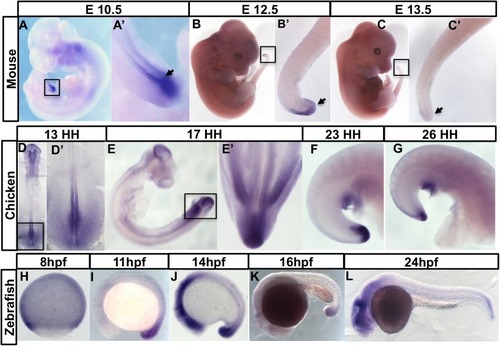
The timing of axial Greb1 expression is coincidentwith axial elongation in vertebrate embryos. (A–D) Mouse embryos and their tail regions at different embryonic stages. (A,A′) Dorsal view of E8.5 embryo showing expression in the caudal lateral epiblast (CLE). PS: primitive streak. (B,B′) Lateral view of E10.5 embryo, showing the Greb1-expressing tail region. (C) Lateral view of E12.5 tail region, showing reduced Greb1 expression. (D) Lateral view of E13.5 tail region, showing that expression is lost. (E–F) Greb1 expression in chick embryos at different stages: (E,E′) dorsal views of HH13 embryo and its tail region; (F) lateral and (F′) ventral view of a tail region at HH17; (G) lateral view of HH26 embryo, showing that Greb1 expression in the tailbud is almost gone. H–L are lateral views of zebrafish embryos of the indicated ages (hpf, hours post-fertilisation). Each pattern was analysed in two independent experiments using, for each stage, at least five mouse, or 10–15 chicken or zebrafish embryos. Tailbud regions are arrowed. Boxes in lower magnification images show the tail regions with magnified views in the adjacent panel.
|
