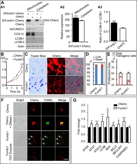
DrFundc1 reduced cell viability while inducing autophagy and apoptosis in transgenic 293 T cells. (A) Western blotting of proteins extracted from mitochondria and cytoplasm from transgenic 293 T cells transfected with pCS2 + -Drfundc1-Cherry-His as well as pCS2 + -Cherry plasmids. Antibodies used were anti-Cherry (detecting Drfundc1-Cherry-His and Cherry), anti-FUNDC1, anti-COX IV, anti-LC3B, and anti-β-ACTIN. (A1) Results of Western blotting. (A2) Gray-scale analyses of DrFundc1 using ImageJ in mitochondria and cytoplasm of cells transfected with pCS2 + -Drfundc1-Cherry-His. (A3) LC3B-II:LC3B-I ratio in mitochondria of transgenic cells transfected with pCS2 + -Drfundc1-Cherry-His and pCS2 + -Cherry. (B) DrFundc1 decreased viability of 293 T cells, measured by MTT assay. (C-D) DrFundc1 increased 293 T cell mortality, measured by Trypan Blue staining. (E) DrFundc1 decreased cell proliferation in transgenic cells, detected using BrdU incorporation. (F) DrFundc1 led to apoptosis of 293 T cells, detected using TUNEL assay. Arrows show apoptotic cells. Red indicates DrFundc1-Cherry or Cherry, while green indicates TUNEL-positive cells. Positive control: Cherry + DNase I. Negative control: incubation without TdT enzyme. (G) Expressional fold change of autophagy- and apoptosis-related genes, detected using qRT-PCR. β-ACTIN was used as an internal control. Significant differences between cells transfected with different plasmids are shown as asterisks. *P < 0.05; **P < 0.01; ***P < 0.001.
|
