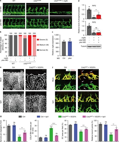
PIP3 exhaustion governs VEGFA-induced regression of CDS2-deficient endothelium. Confocal images (a) and quantitative analysis (b) of vessel-deficient phenotype in WT zebrafish embryos or cds2 mutants with or w/o vegfa OE, with control MO or pten MO (combination of ptena and ptenb MO) injection. The counted ISV number shown on the top (b) is from 15–20 embryos per group. c Relative vegfa mRNA level of cds2 mutants with vegfa OE injected with ctrl or pten MO. The expression was normalized to WT embryos. n = 3 samples per group, 15–20 embryos pooled for each sample. dpten knockdown partially restored PIP3, but not PIP2 level in cds2-deficient endothelium with vegfa OE. Western blotting analysis on a-Tubulin serves as the internal control for cell amounts in each sample collected during lipid quantitation analysis. n = 4 samples each group. Confocal images (e and f) and quantitative analysis (g) of P7 retinal vessels stained by IB4 (e) and IB4/COL4 (f) from control or VEGFA-injected Cds2iΔEC mice treated with bpV (PTEN inhibitor) or vehicle (saline). bpV (2 mg kg−1) or vehicle was given three times at P2, P4 and P6. Arrows show regressed vessels. Angiogenic sprouts, COL4+/IB4− empty sleeves, endothelial area and branch points were quantified in (g), n = 6–10 mice per group. Scale bars, 100 μm (a and f) and 200 μm (e). Error bars, mean ± SEM. *P < 0.05; **P < 0.01; ***P < 0.001; ****P < 0.0001; ns, not significant (P ≥ 0.05). See also Supplementary information, Fig. S6
|
