
Fig. 3
- ID
- ZDB-FIG-191210-3
- Publication
- Sagarin et al., 2019 - Anterior Trunk Muscle Shows Mix of Axial and Appendicular Developmental Patterns
- Other Figures
- All Figure Page
- Back to All Figure Page
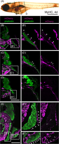
The PHM has superficial and internal mCherry?positive connective tissue cells. A, Whole?mount MyHC stain showing positions of sections shown in B?D. B?D, Transverse sections of regions indicated in A of Tg(drl:creERT2);Tg(ubi:Switch) embryos, labeled for mCherry (magenta) and phalloidin (green). mCherry expressing cells are derived from lateral plate mesoderm. B??E?, magnifications of boxed regions in B?E. B?B?, Section at the level of the anterior PHM. mCherry?expressing cells are found along the entire superior surface of the PHM (arrowheads). C?C?, Section through more posterior PHM. Weak mCherry expression is seen along a portion of the superior surface of the PHM (arrowheads). D?D?, Section through trunk myotome. mCherry expression is absent from the surface of the trunk myotome. E?E?, Transverse section at the level of the anterior PHM in a 9.3?mm juvenile. mCherry positive cells are found along the entire superficial surface of the PHM (closed arrowheads). mCherry expression is present between muscle fibers in the PHM and PFM (open arrowheads). Scale bar in A is 1?mm. Scale bar in D, for B?D, is 100??m. Scale bar in E is 500??m |