
Fig. 5
- ID
- ZDB-FIG-190910-25
- Publication
- O'Connor et al., 2019 - Modulation of Agrin and RhoA Pathways Ameliorates Movement Defects and Synapse Morphology in MYO9A-Depleted Zebrafish
- Other Figures
- All Figure Page
- Back to All Figure Page
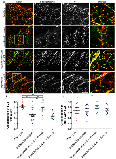
NMJ morphology of myo9aa/ab crispant zebrafish at 120 hpf. (A) Representative images of NMJs in wild-type, myo9aa/ab crispant, and NT1654 (0.15 ng) or fasudil-treated (10 µm) crispant zebrafish at 120 hpf. Acetylcholine receptors stained with αBTx (green), and motor neurons detected with an antibody against SV2 (red). White boxes demark areas enlarged in the right-hand panel. Scale bar = 50 µm. (B) Colocalisation of SV2-positive signal with αBTx and (C) colocalisation of αBTx with SV2-positive signal using Mander’s correlation coefficient (0 = no colocalisation, 1 = full colocalisation). Wildtype (n = 6 fish), myo9aa/ab crispant (n = 12 fish), crispant treated with NT1654 (0.15 ng, n = 8 fish), and crispant treated with fasudil (10 µm, n = 11 fish) were subject to analysis. Error bars = mean ± S.E.M. ** p ≤ 0.01 and ns = not significant, One-way ANOVA. |
| Fish: | |
|---|---|
| Conditions: | |
| Knockdown Reagents: | |
| Observed In: | |
| Stage: | Day 5 |