|
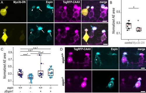
In the absence of Espin actin-bundling activity, CSF-cNs form shorter apical extensions.(A) Z-projections of whole-mounted spinal cords at 72 hpf showing mosaic expression of Myo3b-DN under the control of the pkd2l1 promoter. Immunostaining reveals Espin (cyan) in TagRFP-CAAX-positive CSF-cNs (magenta) expressing Myo3b-DN (yellow, arrowheads) or not (arrows). In ventral CSF-cNs expressing Myo3b-DN, Espin staining is reduced (observed in 9 cells out of 9), and the AE appears smaller compared with wild-type cells. (B) Quantification of the normalized area covered by the AE of ventral cells expressing Myo3b-DN (N = 9 cells) compared with nonexpressing neighboring cells (control; N = 10 cells). Cells expressing Myo3b-DN form a significantly smaller AE (N = 5 fish; p = 0.025). (C) The same quantification was performed in espin−/− ventral cells at 72 hpf (N = 30 cells in 5 fish) compared with wild-type cells (N = 16 cells in 5 fish). Mutant cells display significantly smaller AEs (p1 = 8.4010 × 10⁻⁶). In wild-type ventral cells, the overexpression of the zEspin1 was sometimes associated with abnormally long microvilli (observed in 2 cells out of 9; red chevrons; p2 = 0.2123). In espin−/− cells, the mutant phenotype was rescued by zEspin1 (N = 15 cells; p3 = 0.3029 and p4 = 3.8556 × 10⁻⁶). (D) Z-projections of whole-mounted spinal cords at 72 hpf showing mosaic expression of zEspin1 under the control of the pkd2l1 promoter. Immunostaining for Espin (cyan) in TagRFP-CAAX-positive ventral CSF-cNs (magenta) reveals the loss of Espin immunoreactivity in espin−/− larvae, which is retrieved in mutant cells expressing zEspin1 (nuclear RFP; magenta; stars). Scale bars, 5 μm. Underlying data can be found in S1 Data. AE, apical extension; CSF-cN, cerebrospinal fluid-contacting neuron; DN, dominant-negative; hpf, hours post fertilization; Myo3b, myosin 3b;n.s., not significant; RFP, red fluorescent protein; zEspin1, zebrafish Espin isoform 1.
|