
Fig. 5
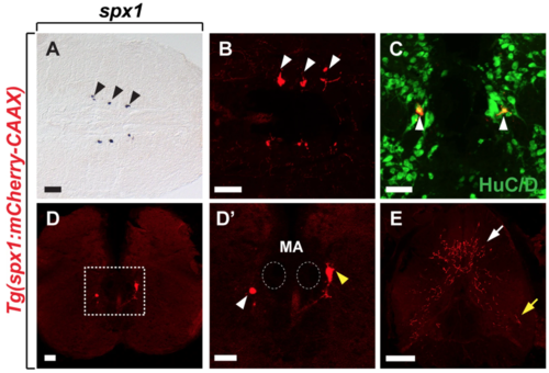
Characterisation of SPX1 neurons in the hindbrain of adult zebrafish. Horizontal (A,B) and transverse (C–E) sections of the brain (A–D’) and spinal cord (E) of adult Tg(spx1:mCherry-CAAX) zebrafish. (A,B) Anterior is to the left and (C–E) dorsal is to the top. SPX1 neurons revealed by whole-mount in situ RNA hybridization with spx1 RNA (A) and spx1:mCherry expression (B). (C) Labelling of the hindbrain of Tg(spx1:mCherry-CAAX) adult zebrafish with anti-HuC/D antibody. (B,C) White arrowheads indicate spx1:mCherry neurons in the brainstem. (D) spx1:mCherry expression is detected in the inferior reticular formation region of the hindbrain. (D’) Magnified image of the boxed area in the panel D. Dotted circles mark the position of Mauthner axons in the hindbrain. White and yellow arrowheads indicate the inferior reticular formation region and vagal motor nucleus region, respectively. (E) Axons of hindbrain SPX1 neurons projecting to the spinal cord. White and yellow arrows indicate axonal projections in the dorsal and ventral spinal cord, respectively. Abbreviation: MA, Mauthner axon. Scale bar: 50 μm. |
| Genes: | |
|---|---|
| Antibody: | |
| Fish: | |
| Anatomical Terms: | |
| Stage: | Adult |