
FIGURE
Fig. 3-S2
- ID
- ZDB-FIG-190201-11
- Publication
- Naylor et al., 2018 - A novel mechanism of gland formation in zebrafish involving transdifferentiation of renal epithelial cells and live cell extrusion
- Other Figures
- All Figure Page
- Back to All Figure Page
Fig. 3-S2
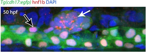
Hnf1b nuclear export at 50 hpf. Lateral view of a 50 hpf Tg(cdh17:egfp) (green) embryo immunostained for Hnf1b (red) and counterstained for DAPI (blue). Hnf1b in the CS is largely non-nuclear (white arrow), in contrast to nuclear Hnf1b in the tubules (black arrow). |
Expression Data
| Gene: | |
|---|---|
| Antibody: | |
| Fish: | |
| Anatomical Terms: | |
| Stage: | Long-pec |
Expression Detail
Antibody Labeling
Phenotype Data
Phenotype Detail
Acknowledgments
This image is the copyrighted work of the attributed author or publisher, and
ZFIN has permission only to display this image to its users.
Additional permissions should be obtained from the applicable author or publisher of the image.
Full text @ Elife