FIGURE
Fig. 6
- ID
- ZDB-FIG-180912-19
- Publication
- Kinoshita et al., 2018 - Functional roles of the Ripply-mediated suppression of segmentation gene expression at the anterior presomitic mesoderm in zebrafish
- Other Figures
- All Figure Page
- Back to All Figure Page
Fig. 6
|
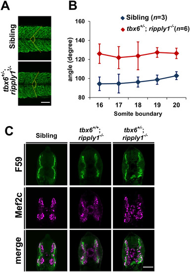
Myogenesis is perturbed in tbx6+/?; ripply1?/? mutants. (A) A myotome stained with MF20 antibody at 36 hpf. The yellow-dashed line was drawn to emphasize the segment boundary and the chevron angle in sibling and tbx6+/?; ripply1?/? embryos. (B) Graph showing the chevron angles in 16th to 20th segments in sibling and tbx6+/?; ripply1?/? embryos. (C) Transverse sections stained with anti-Mef2c and F59 antibodies. Zebrafish body corresponding to the 16th to 20th myotomes at 24 hpf was used for analysis. The signal around the notochord is non-specific signal. Scale bar is 100??m. |
Expression Data
| Antibodies: | |
|---|---|
| Fish: | |
| Anatomical Terms: | |
| Stage Range: | Prim-5 to Prim-25 |
Expression Detail
Antibody Labeling
Phenotype Data
| Fish: | |
|---|---|
| Observed In: | |
| Stage Range: | Prim-5 to Prim-25 |
Phenotype Detail
Acknowledgments
This image is the copyrighted work of the attributed author or publisher, and
ZFIN has permission only to display this image to its users.
Additional permissions should be obtained from the applicable author or publisher of the image.
Reprinted from Mechanisms of Development, 152, Kinoshita, H., Ohgane, N., Fujino, Y., Yabe, T., Ovara, H., Yokota, D., Izuka, A., Kage, D., Yamasu, K., Takada, S., Kawamura, A., Functional roles of the Ripply-mediated suppression of segmentation gene expression at the anterior presomitic mesoderm in zebrafish, 21-31, Copyright (2018) with permission from Elsevier. Full text @ Mech. Dev.

